, 25µg")
- Remove this product from my favorite's list.
- Add this product to my list of favorites.
Products

Newsletter
 |  |  |  |  |  |
Background
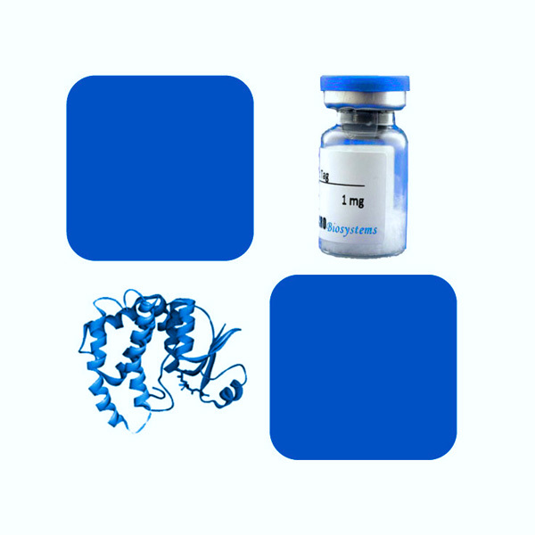
Programmed cell death protein 1 (PD-1) is also known as CD279 and PDCD1, is a type I membrane protein and is a member of the extended CD28/CTLA-4 family of T cell regulators. PDCD1 is expressed on the surface of activated T cells, B cells, macrophages, myeloid cells and a subset of thymocytes. PD-1 has two ligands, PD-L1 and PD-L2, which are members of the B7 family. PD-L1 is expressed on almost all murine tumor cell lines, including PA1 myeloma, P815 mastocytoma, and B16 melanoma upon treatment with IFN-γ. PD-L2 expression is more restricted and is expressed mainly by DCs and a few tumor lines. PD1 inhibits the T-cell proliferation and production of related cytokines including IL-1, IL-4, IL-10 and IFN-γ by suppressing the activation and transduction of PI3K/AKT pathway. In addition, coligation of PD1 inhibits BCR-mediating signal by dephosphorylating key signal transducer. In vitro, treatment of anti-CD3 stimulated T cells with PD-L1-Ig results in reduced T cell proliferation and IFN-γ secretion. Monoclonal antibodies targeting PD-1 that boost the immune system are being developed for the treatment of cancer.
Source
Recombinant Biotinylated Human PD-1, Fc,Avitag (PD1-H82F1) is expressed from human 293 cells (HEK293). It contains AA Leu 25 - Gln 167 (Accession # Q15116-1).
Predicted N-terminus: Leu 25
Molecular Characterization
This protein carries a human IgG1 Fc tag at the C-terminus, followed by an Avi tag (Avitag™).
The protein has a calculated MW of 44.4 kDa. The protein migrates as 55-66 kDa when calibrated against Star Ribbon Pre-stained Protein Marker under reducing (R) condition (SDS-PAGE) due to glycosylation.
Biotinylation
Biotinylation of this product is performed using Avitag™ technology. Briefly, the single lysine residue in the Avitag is enzymatically labeled with biotin.
Biotin:Protein Ratio
Passed as determined by the HABA assay / binding ELISA.
Endotoxin
Less than 1.0 EU per μg by the LAL method.
Purity
>95% as determined by SDS-PAGE.
>95% as determined by SEC-MALS.
Formulation
Lyophilized from 0.22 μm filtered solution in PBS, pH7.4. Normally trehalose is added as protectant before lyophilization.
Reconstitution
Please see Certificate of Analysis for specific instructions.
For best performance, we strongly recommend you to follow the reconstitution protocol provided in the CoA.
Storage
For long term storage, the product should be stored at lyophilized state at -20°C or lower.
Please avoid repeated freeze-thaw cycles.
This product is stable after storage at:
-20°C to -70°C for 12 months in lyophilized state;
-70°C for 3 months under sterile conditions after reconstitution.
Bioactivity
Please refer to product data sheet.
(1) "B.infantis enhances immunotherapy for Guillain-Barre syndrome through regulating PD-1"
Shi, Nian, Qu et al
BMC Neurol (2023) 23 (1), 48
(2) "[Single-center study of different treatment for advanced or unresectable angiosarcoma patients]"
Peng, Xu, Liu et al
Zhonghua Zhong Liu Za Zhi (2023) 45 (1), 74-81
(3) "Identification of a tumour immune barrier in the HCC microenvironment that determines the efficacy of immunotherapy"
Liu, Xun, Ma et al
J Hepatol (2023)
Showing 1-3 of 26040 papers.
Recombinant Human PD-L1/B7-H1 Protein, low endotoxin, Fc Tag, 100µg - 429,00 €
Recombinant Mouse PD-L1 /CD274 /B7-H1 Protein, With C-Fc Tag, 200µg - 364,00 €
Recombinant Cynomolgus / Rhesus macaque PD-L1 / B7-H1 Protein, His tag, 100µg - 468,00 €
Recombinant Mouse PD-L1 /CD274 /B7-H1 Protein, His tag, 100µg - 435,50 €
Recombinant Human PD-L1 / B7-H1 Protein, mouse IgG1 Fc Tag, low endotoxin (HPLC-verified), 100µg - 403,00 €


Follow us